Background: The coexistence of neuromyelitis optica spectrum disorder (NMOSD) and longitudinally extensive transverse myelitis (LETM) in tuberculosis (TB) is rare and presents a diagnostic challenge.
Case Presentation: We report the case of a 39-year-old man receiving anti tuberculous treatment for smear-positive pulmonary tuberculosis (PT) for one week. He presented with acute left-sided weakness that rapidly progressed to quadriparesis within 24 hours, accompanied by a T2 sensory level and sphincter dysfunction. Cervical spine MRI demonstrated longitudinally extensive transverse myelitis (LETM) from C3 to C7, while brain MRI showed features suggestive of tuberculous meningitis (TBM). Cerebrospinal fluid analysis revealed lymphocytic pleocytosis and elevated protein. Urinary tuberculosis was also confirmed by positive urine acid-fast bacilli testing in the setting of renal tubular electrolyte wasting. Based on these findings, the initial diagnosis was disseminated tuberculosis involving the pulmonary, urinary, central nervous systems, with LETM and meningitis. Subsequently, he developed sudden bilateral blindness, and fundoscopy demonstrated bilateral optic atrophy. Serum aquaporin-4 (AQP4) antibody was weakly positive, supporting concomitant AQP4-IgG-positive neuromyelitis optica spectrum disorder (NMOSD). Anti-tuberculous therapy and high-dose intravenous methylprednisolone were commenced. Pulmonary symptoms improved, but neurological and visual recovery remained limited.
Conclusion: This case highlights the diagnostic complexity of NMOSD occurring in the setting of disseminated tuberculosis. Tuberculosis may mimic or coexist with NMOSD, and careful evaluation for active infection is essential before initiating immunosuppressive therapy.
Keywords: tuberculosis; NMOSD; longitudinally extensive transverse myelitis; optic atrophy; aquaporin-4 antibody.
Introduction
Tuberculosis involving the spinal cord is associated with high mortality and disabling long term outcomes. Despite the widespread TB burden in low and middle income countries, large scale data on tuberculous spinal cord involvement is lacking 1. Tuberculous myelopathy in the absence of Pott’s disease comprises all atypical forms of spinal tuberculosis such as tubercular myelitis, intradural tuberculosis and spinal cord complications of tubercular meningitis.Nearly half of the patients with TBM may concomitantly harbor spinal cord and spinal nerve root (Primary spinal cord TB). Secondary spinal cord involvement may also occur due to vertebral TB 2.
Neuromyelitis optica spectrum disorder (NMOSD) is an autoimmune inflammatory disease of the central nervous system, with an estimated adult prevalence of 0.34–10 per 100,0003. Most patients have serum anti-aquaporin-4 immunoglobulin G (anti-AQP4-IgG) antibodies, a highly specific disease marker that targets AQP4 on astrocytic end-feet and activates complement 4. Several reports have described coexistence of NMOSD and CNS TB ,however, the pathophysiological relationship between them remains unclear 5,6.
Case report
Clinical presentation
A 39-year-old married man, a Buddhist container truck driver from Insein, was admitted to Yangon General Hospital on June 2026 with a one-day history of sudden onset left-sided weakness. He denied dysphagia, facial asymmetry, dysarthria, headache, dizziness, vomiting, seizures, or loss of consciousness. On examination, he was fully conscious with Glasgow Coma Score of 15/15. His vital signs were stable and there was no cranial nerve palsy. Muscle power was 4/5 on the left side, with reduced muscle tone and deep tendon reflexes. Clonus was absent while the plantar response was extensor on the left, flexor on the right.
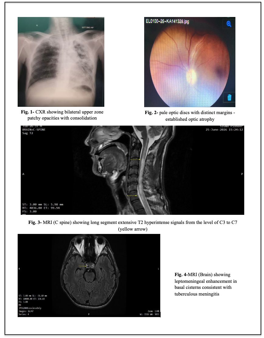
He had no previous history of hypertension, diabetes, or previous stroke, however he was a chronic smoker and alcohol user. On further questioning, he reported one-month history of intermittent productive cough with white sputum and evening rise of temperature. Chest radiograph showed bilateral upper zone patchy opacities with areas of consolidation (Figure 1). Ziehl-Neelsen staining of the sputum revealed acid-fast bacillus (AFB), suggestive of Pulmonary Tuberculosis. He had been started on anti-tuberculous treatment one week before admission.
A working diagnosis of cerebrovascular accident was initially made, However, in view of the confirmed pulmonary tuberculosis and the neurological presentation, tuberculoma, tuberculous meningitis, and tuberculous vasculitis, were also considered as differential diagnosis.
Initial Investigations
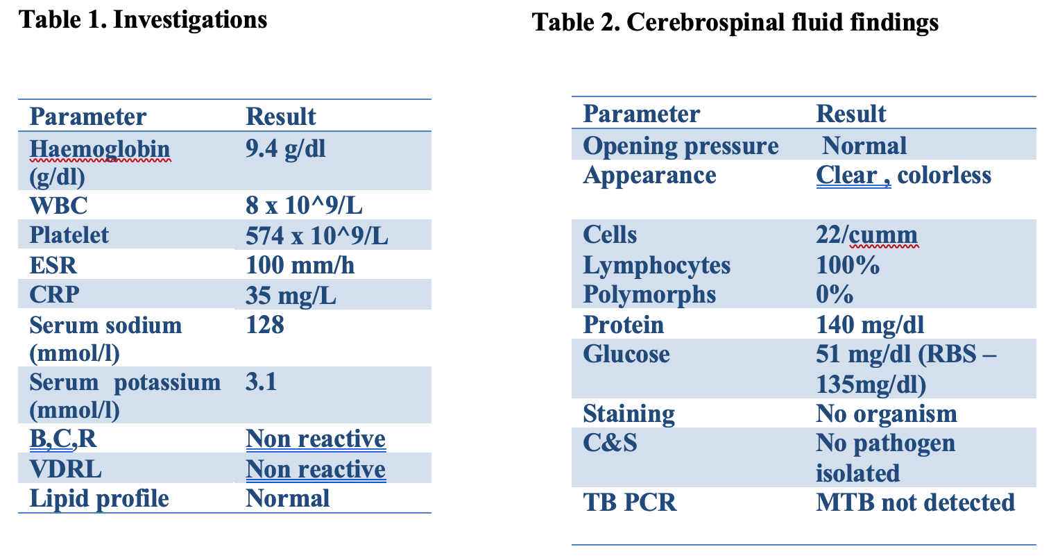
Electrocardiogram showed normal sinus rhythm, random blood sugar of 166 mg/dl, and non-contrast computed tomography of the head showed no significant abnormality. Initial laboratory investigations are presented in Table 1.
Clinical Course
On the second day after admission, weakness progressed to involve the right side. Tone was reduced in all four limbs. Muscle power was 2/5 in the right upper and lower limbs, 0/5 in the left upper limb, and 1/5 in the left lower limb. Deep tendon reflexes were diminished, while bilateral plantar responses were extensor. A sensory level was identified at T2. The working diagnosis was therefore revised to quadriparesis with sphincter involvement and a T2 sensory level, most consistent with transverse myelitis (TM).
On further questioning, it became apparent that the patient had experienced sudden painless vision loss two days after quadriparesis. although he had not volunteered this symptom previously. Fundoscopic examination demonstrated bilateral pale optic discs with distinct margins consistent with established optic atrophy. There was no cherry-red spot, exudate, haemorrhage, papilloedema and choroidal tubercles (Fig. 2).
Diagnostic Workup and Evaluation
MRI of the cervical spine showed a lesion extending from C3 to C7 involving three or more contiguous vertebral segments. This was characteristic of longitudinally extensive transverse myelitis (Fig. 3). CSF examination showed lymphocytic pleocytosis and elevated protein. The complete CSF findings are presented in Table 2.
MRI of the brain showed leptomeningeal enhancement in bilateral basal cistern, compatible with tuberculous meningitis (Figure 4). Autoimmune screening was negative for antinuclear antibody, extractable nuclear antigen profile and rheumatoid factor.
The coexistence of longitudinally extensive transverse myelitis and bilateral optic nerve involvement raised suspicion for NMOSD. Subsequently, serum anti-aquaporin-4 antibody was weakly positive. Myelin oligodendrocyte glycoprotein (MOG) antibody was found out to be negative
Clinical course and complications
Antituberculous therapy was continued alongside intravenous dexamethasone. After three weeks of antituberculous treatment, and following consultation with the Neuromedical department. intravenous high-dose methylprednisolone was administered. Although the pulmonary symptoms improved, there was no corresponding neurological recovery.
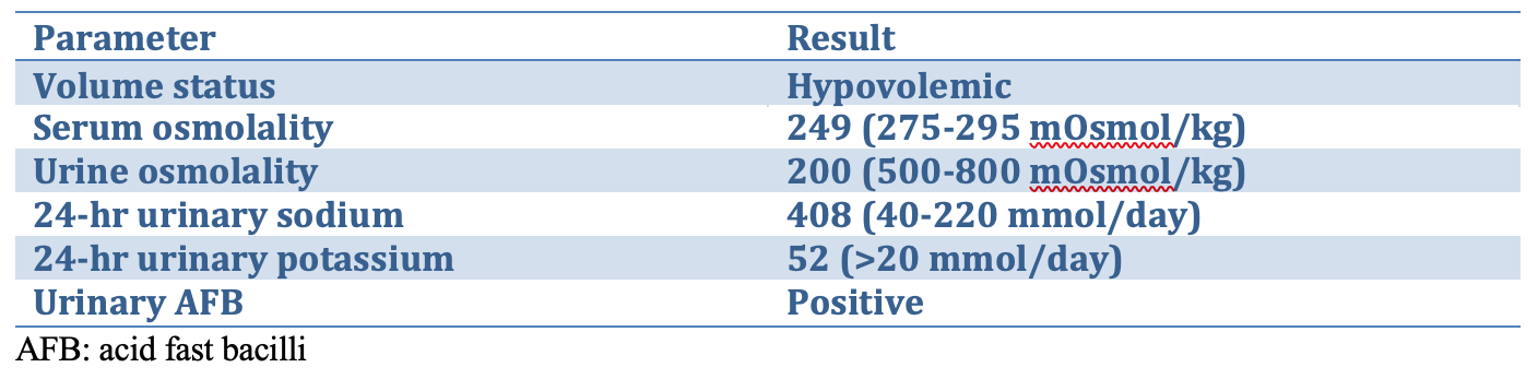
During this period, the patient’s level of consciousness gradually deteriorated despite high-dose dexamethasone and antituberculous therapy. Repeat assessment revealed profound hyponatraemia (Na 119 mmol/L) accompanied by the evidence of tubular electrolyte loss (Table 3). Subsequent urine testing was positive for acid-fast bacilli confirming genitourinary tuberculosis. The final diagnosis was therefore disseminated tuberculosis involving the pulmonary, genitourinary systems, with longitudinally extensive transverse myelitis (LETM), occuring concomitant with neuromyelitis optica spectrum disorder (NMOSD).
The prognosis remains guarded. The severity of the neurological presentation, particularly the development of spastic quadriplegia, places the patient at substantial risk of permanent neurological disability. Similarly, the development of optic atrophy makes meaningful visual recovery considerably less likely, as established axonal damage is irreversible. Nevertheless, the patient has survived a severe and complex illness with continued support from his family.
Table 3-Urinary osmolarity and electrolytes
Discussion
Tuberculous spinal cord involvement is rare and may present as either short-segment myelitis or LETM, even without vertebral disease. Proposed mechanisms include venous congestion related to tuberculous meningitis, vasculitis or thrombosis causing ischaemic injury, intramedullary tuberculoma, and direct cord infarction. The cervicodorsal region appears to be most frequently affected 2. Accordingly, tuberculosis should remain in the differential diagnosis of myelitis in endemic settings, as LETM is not specific to NMOSD and may occur in CNS tuberculosis 7.
The association between tuberculosis and AQP4-IgG-positive NMOSD has been reported only rarely, mainly in case reports and small series, and causality remains unproven. Reports from Mexico described patients with tuberculous meningitis who subsequently developed AQP4-antibody-positive LETM and improved with anti tuberculous therapy and corticosteroids 8.
Similarly, a Chinese series included one patient with pulmonary tuberculosis, AQP4-IgG-positive NMOSD, and another with tuberculosis-associated LETM without detectable AQP4-IgG5. Other reports have described fatal or clinically typical NMOSD occurring alongside pulmonary or renal tuberculosis. 9,10 These cases highlight the difficulty of distinguishing infection-related myelitis from coincident autoimmune disease.
Infections may act as environmental triggers for autoimmune disease, although the underlying mechanisms remain uncertain. In a study of 19 AQP4-IgG-positive patients, Koga et al. found serological evidence of recent viral infection in some patients assessed during the acute phase, supporting a possible association without establishing causation 11. Molecular mimicry, epitope spreading, and secondary immune amplification have been proposed. Experimental studies have also shown that antibodies against mycobacterial mannan can affect blood–brain barrier integrity; however, direct molecular mimicry between Mycobacterium tuberculosis and aquaporin-4 has not been demonstrated in humans 12.
In our patient, the combination of LETM, bilateral optic involvement with established optic atrophy, and AQP4-IgG seropositivity supports concomitant NMOSD rather than tuberculosis-associated myelitis alone. Nevertheless, disseminated tuberculosis may have contributed to or triggered the neurological disease, although this cannot be established from a single case.
Management is challenging because NMOSD requires immunosuppression, whereas corticosteroids and other immunosuppressive therapies may promote tuberculosis reactivation or dissemination. In this case, antituberculosis therapy was continued alongside corticosteroids, with careful monitoring for infectious complications.
Ethambutol warrants particular caution in patients with visual symptoms because of its recognised optic toxicity. In this patient, bilateral sudden painless visual loss developed five days after initiation of antituberculous therapy and subsequently progressed to established optic atrophy. Although the visual deterioration was temporally associated with the initiation of anti tuberculous treatment, the underlying cause of the optic neuropathy was considered in the context of concomitant NMOSD and disseminated tuberculosis. Given the potential for further optic nerve injury, ethambutol was substituted with levofloxacin. However, the presence of established axonal loss limits the likelihood of meaningful visual recovery.
Conclusion
Diagnosis and management of patients with spinal cord tuberculosis are difficult and prognosis is striking poor. Tuberculosis-associated neurological manifestations and NMOSD may coexist, creating a diagnostic challenge in distinguishing tuberculosis-related myelitis from an autoimmune demyelinating disorder. Clinicians in tuberculosis-endemic settings should remain alert to such diagnostic overlap and consider the overall clinical picture when establishing the diagnosis.
Disclosure
The authors declare no conflict of interest.
References
- Sharma SK, Ryan H, Khaparde S, Sachdeva KS, Singh AD, Mohan A, et al. (2017) Index-TB guidelines: guidelines on extrapulmonary tuberculosis for India. Indian J Med Res. 2017;145:448-63.
- Garg D, Radhakrishnan DM, Agrawal U, Vanjare HA, Gandham EJ, Manesh A. (2023). Tuberculosis of the spinal cord. Ann Indian Acad Neurol. 26(2):112-26. doi:10.4103/aian.aian_578_22.
- Papp V, Magyari M, Aktas O, Berger T, Broadley SA, Cabre P, et al. (2021). Worldwide incidence and prevalence of neuromyelitis optica: a systematic review. Neurology. 96(2):59-77. doi:10.1212/WNL.0000000000011153.
- Wingerchuk DM, Banwell B, Bennett JL, Cabre P, Carroll W, Chitnis T, et al. (2015). International consensus diagnostic criteria for neuromyelitis optica spectrum disorders. Neurology. 85(2):177-89.
- Zhang Y, Zhu M, Wang L, Shi M, Deng H. (2018). Longitudinally extensive transverse myelitis with pulmonary tuberculosis: two case reports. Medicine (Baltimore). 97(3):e9676.
- De Saráchaga AJ, Rivera-Chávez LF, López-Martínez MS. (2020). Neuromyelitis optica spectrum disorder (NMOSD) with hypothalamic involvement and central nervous system tuberculosis: a case report. Clin Neurol Neurosurg. 193:105751.
- Trebst C, Raab P, Voss EV, et al. (2011). Longitudinal extensive transverse myelitis—it’s not all neuromyelitis optica. Nat Rev Neurol. 7(12):688-98.
- Gomez-Figueroa E, Garcia-Estrada C, Casallas-Vanegas A, Zabala-Angeles I, Lopez-Martinez R, Corona-Vazquez T, et al. (2020). Anti-aquaporin-4 antibody-positive neuromyelitis optica spectrum disorder associated with central nervous system tuberculosis. Clin Exp Neuroimmunol. 11(1):40-4. doi:10.1111/cen3.12548.
- Sridhar S, Chan JFW, Yuen KY. (2014). Fatal anti-aquaporin-4 seropositive neuromyelitis optica spectrum disorder in tuberculosis. BMC Infect Dis. 14:470.
- Saied Z, Nabli F, Rachdi A, et al. (2021). Neuromyelitis optica and concomitant pulmonary tuberculosis: a case report. J Med Case Rep. 15:391.
- Koga M, Takahashi T, Kawai M, Fujihara K, Kanda T. (2011). A serological analysis of viral and bacterial infections associated with neuromyelitis optica. J Neurol Sci. 300(1-2):19-22. doi:10.1016/j.jns.2010.10.013.
- Namer IJ, Steibel J. (2000). Antibody directed against mannan of the Mycobacterium tuberculosis cell envelope provokes blood-brain barrier breakdown. J Neuroimmunol. 103:63-8.
Author Information
May-Kyi-Oo, Ni-Ni-Aung, Min-Zaw-Oo
- Senior Consultant physician, Department of Medicine, Yangon General Hospital, Yangon
- Junior consultant physician, Department of Medicine, Yangon General Hospital, Yangon
- Professor, Department of Medicine, Yangon General Hospital, Yangon