Abstract
Malignant pleural effusion (MPE) is a common manifestation of advanced malignancy and is associated with substantial symptom burden, impaired quality of life and poor prognosis.
Contemporary management has evolved considerably, with increasing emphasis on ambulatory care, minimally invasive interventions and patient-centred decision-making.
This review provides a practical overview of the investigation and management of MPE based on current evidence and guideline recommendations. Diagnostic approaches, including thoracic ultrasound, pleural fluid cytology and pleural biopsy, are discussed. Therapeutic strategies, including therapeutic pleural aspiration, talc pleurodesis, indwelling pleural catheters (IPCs) and thoracoscopic approaches, are reviewed alongside evidence from landmark trials that have shaped modern practice. Special clinical situations, including non-expandable lung and septated MPE, are also considered, together with the complementary role of systemic anti-cancer therapy.
No single intervention is universally appropriate for all patients with MPE. Management should therefore be individualised according to symptom burden, lung expandability, prognosis, anticipated treatment burden and patient preference. While contemporary guidelines provide an evidence-based framework for care, their application should be adapted pragmatically to local expertise and healthcare resources. Ultimately, the goals of MPE management remain unchanged: to alleviate symptoms, minimise unnecessary interventions and optimise quality of life through shared decision-making and patient-centred care.
Introduction
Malignant pleural effusion (MPE) is a common complication of advanced malignancy, affecting an estimated 1 million people globally each year, with approximately 50,000 new cases diagnosed annually in the UK. ¹ It is associated with impaired functional status, reduced quality of life and substantial symptom burden. Lung and breast cancers are the leading causes of MPE, together accounting for approximately 50–65% of cases, while lymphoma, ovarian malignancy and malignant pleural mesothelioma are also important causes. ¹
The development of MPE generally reflects advanced disease and is associated with poor prognosis, with median survival ranging from 3 to 12 months depending on tumour type and patient factors. ¹ The primary aim of MPE management is therefore palliative, focusing on effective symptom control while minimising treatment burden and hospitalisation. Over the past decade, pleural medicine has evolved considerably with increasing adoption of ambulatory care pathways, minimally invasive interventions and indwelling pleural catheters (IPCs). Contemporary management increasingly emphasises the selection of interventions according to symptom burden, lung expandability, anticipated prognosis and patient preference. ¹–³
International guidelines have incorporated evidence from landmark randomised controlled trials and observational studies, resulting in important changes in clinical practice. ²˒³ This review provides a practical overview of the investigation and management of MPE based on current literature and guideline recommendations. ¹–³
Clinical Presentation
Patients with MPE commonly present with progressive dyspnoea, non-productive cough and chest discomfort. Symptoms often develop gradually and reflect pleural fluid accumulation, underlying lung compression and the extent of the underlying malignancy. Constitutional symptoms, including fatigue, anorexia and weight loss, may also be present. ¹–³
Clinical examination findings vary according to the size of the effusion and may include reduced breath sounds, dullness to percussion and reduced chest expansion on the affected side. Large pleural effusions may occasionally result in mediastinal shift and significant respiratory compromise. ²˒³
Importantly, the severity of breathlessness does not always correlate with the volume of pleural fluid. Symptoms may also be influenced by underlying cardiopulmonary disease, non-expandable lung (NEL), reduced physiological reserve and concurrent systemic illness. Consequently, decisions regarding pleural intervention should be guided by both clinical assessment and radiological findings rather than imaging appearance alone. ¹˒²
Investigation of Suspected Malignant Pleural Effusion
Chest radiography is usually the initial imaging modality in patients presenting with suspected pleural effusion and may demonstrate unilateral or bilateral pleural fluid accumulation. Thoracic ultrasound (TUS) has become an essential component of pleural assessment and should be used to guide pleural aspiration, improving procedural accuracy and reducing complications. ²˒³
Diagnostic pleural aspiration remains a key initial investigation in patients with unexplained unilateral pleural effusions. Pleural fluid analysis should be guided by the clinical context and commonly includes protein and LDH with paired serum measurements, cytological examination when malignancy is suspected, and microbiological testing when pleural infection is a differential diagnosis. Light’s criteria remain the standard method for differentiating exudative from transudative pleural effusions.²˒⁴
Pleural fluid cytology is recommended as the initial diagnostic test in patients with suspected secondary pleural malignancy, although its sensitivity varies considerably according to tumour subtype. Adenocarcinoma is generally associated with a higher cytological yield, whereas mesothelioma demonstrates substantially lower sensitivity. Consequently, a negative cytology result does not exclude pleural malignancy, and further investigation is warranted when clinical suspicion remains high.⁵
Contrast-enhanced computed tomography (CT) of the thorax is valuable in assessing pleural thickening, pleural nodularity, underlying lung malignancy and metastatic disease. However, negative CT findings do not reliably exclude pleural malignancy.²˒³ PET-CT may be considered in selected patients with suspicious clinical or CT features despite negative histological results, or when invasive sampling is not feasible; routine use is not recommended.²˒⁶
Where pleural fluid cytology is non-diagnostic, further tissue sampling is frequently required to establish a histological diagnosis and guide oncological management.²˒³ Image-guided pleural biopsy and thoracoscopy both have important diagnostic roles depending on local expertise and clinical context. Current guidance favours image-guided or thoracoscopic pleural biopsy over blind pleural biopsy techniques because of superior diagnostic yield.²˒³ Medical thoracoscopy additionally permits direct visualisation of the pleural cavity and facilitates therapeutic interventions, including talc poudrage pleurodesis, in appropriately selected patients.¹˒²
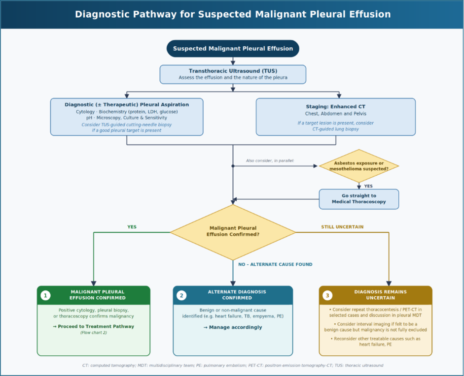
Fig 1. Suggested diagnostic pathway for suspected malignant pleural effusion. Author-generated flowchart informed by the British Thoracic Society Pleural Disease Guideline 2023 and the contemporary review by Piggott et al.¹˒²
Principles of Management
The management of MPE aims to relieve symptoms, prevent recurrent fluid accumulation and minimise treatment burden. Definitive intervention should be considered in patients with recurrent symptomatic effusions, taking into account lung expandability, anticipated prognosis and procedural suitability. ¹–³ In a large retrospective cohort study using US cancer registry-linked Medicare data, 55% of patients required a second pleural procedure following initial thoracentesis; among those with recurrence, 58% required this within 2 weeks. Despite this, only 24% of patients with rapidly recurrent MPE received a guideline-consistent definitive second pleural procedure. ⁷
As several effective treatment options exist, no single intervention is appropriate for all patients. The choice of therapy should therefore be individualised through shared decision-making, considering the expected benefits, potential risks and practical implications of each approach, ensuring that management aligns with patients’ preferences and goals of care.
1. Therapeutic Pleural Aspiration
Therapeutic pleural aspiration provides rapid symptomatic relief and remains an important intervention in MPE. It may be particularly appropriate in patients with limited life expectancy, poor performance status or slowly recurrent effusions. In addition to relieving breathlessness, large-volume pleural aspiration can help determine whether symptoms are attributable to the effusion and assess lung re-expansion before considering definitive pleural intervention. ²˒³
Although aspiration frequently improves symptoms, pleural fluid commonly re-accumulates. In patients with recurrent symptomatic MPE who are likely to benefit from a more durable approach, talc pleurodesis or indwelling pleural catheters (IPCs) may provide more sustained symptom control while reducing the need for repeated procedures and hospital attendance. ¹–³
Therapeutic aspiration carries a small risk of complications, including pneumothorax, bleeding, infection and re-expansion pulmonary oedema, although routine thoracic ultrasound guidance has substantially improved procedural safety. ²˒³
2A

2B
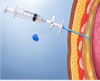
Fig. 2A–B. Therapeutic pleural aspiration. (A) Thoracentesis catheter positioned within the pleural space. (B) Drainage bag attached during therapeutic pleural aspiration.
2. Talc Pleurodesis
Talc pleurodesis remains an established treatment for recurrent symptomatic MPE in patients with expandable lung. By inducing inflammation and adhesion between the visceral and parietal pleura, pleurodesis aims to prevent further pleural fluid accumulation and reduce the need for repeated interventions.¹–³
Pleurodesis may be performed using talc slurry administered through an intercostal chest drain or talc poudrage delivered during thoracoscopy. Current evidence suggests that both approaches are effective, with the TAPPS (Thoracoscopy and Talc Poudrage versus Pleurodesis using Talc Slurry) trial demonstrating similar pleurodesis failure rates between thoracoscopic talc poudrage and talc slurry. ⁸
Successful pleurodesis depends largely on adequate lung re-expansion following pleural drainage. Consequently, patients with non-expandable lung (NEL) are less likely to benefit from this approach, and alternative strategies should be considered.²˒³
Potential disadvantages include inpatient treatment for conventional chest-drain pleurodesis, chest drain-related discomfort and the risk of pleurodesis failure requiring further intervention. Nevertheless, talc pleurodesis remains a valuable treatment option, particularly for patients who prefer to avoid a long-term indwelling catheter and in healthcare settings where community IPC services are limited. Its effectiveness, accessibility and familiarity to clinicians also make it highly relevant across a range of healthcare settings. ¹–³
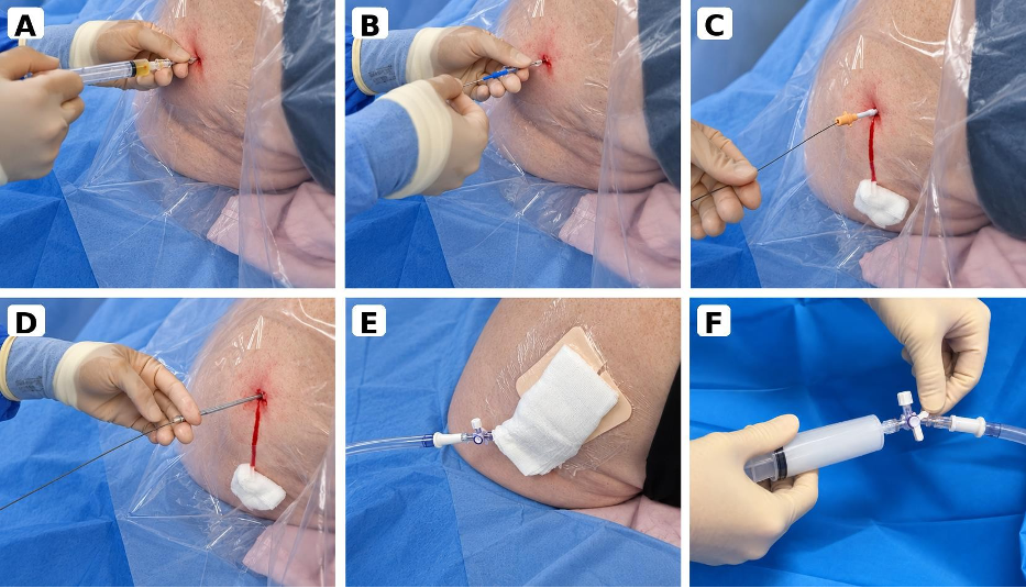
Fig. 3A–F. Seldinger intercostal chest drain insertion and subsequent talc slurry pleurodesis following pleural fluid drainage and confirmation of lung re-expansion. (A) Pleural access with needle aspiration of pleural fluid. (B) Guidewire insertion through the access cannula. (C) Tract dilatation over the guidewire. (D) Advancement of the chest drain using the Seldinger technique. (E) Chest drain in situ and connected to drainage tubing following pleural fluid drainage. (F) Talc slurry administration via the intercostal chest drain following pleural fluid drainage and confirmation of lung re-expansion. Clinical photographs provided by the authors and reproduced with appropriate patient consent.
4A
4B
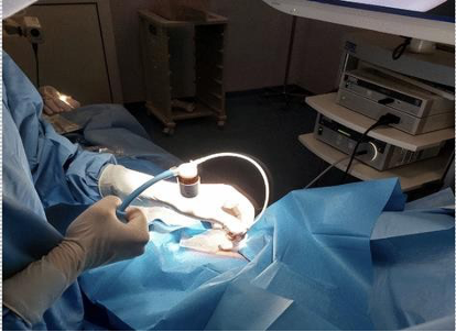
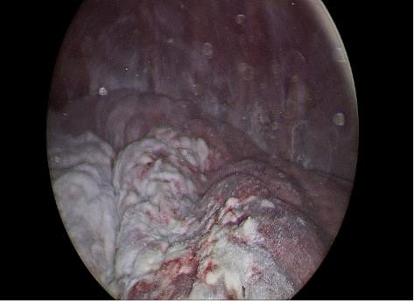
Fig. 4A–B. Talc poudrage during medical thoracoscopy. (A) Insufflation of sterile talc through the dedicated thoracic port. (B) Intrathoracic endoscopic appearance following talc deposition. Reproduced from Fantin et al.⁹ under the terms of the Creative Commons Attribution-NonCommercial 4.0 International License (CC BY-NC 4.0).
3. Indwelling Pleural Catheters (IPC)
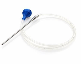
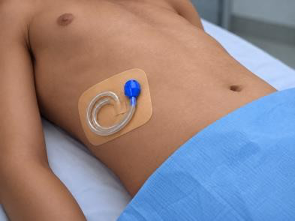
Indwelling pleural catheters (IPCs) have become an established treatment option for recurrent symptomatic MPE. IPCs allow intermittent outpatient drainage of pleural fluid through a tunnelled catheter, facilitating symptom control while reducing the need for repeated pleural procedures and prolonged hospitalisation. ¹–³
IPCs are particularly valuable in patients with non-expandable lung (NEL), failed pleurodesis or a preference for ambulatory management. Contemporary guidelines recognise IPCs as an established first-line definitive treatment option in appropriately selected patients. ²˒³
Randomised controlled trials have demonstrated that IPCs provide symptom relief and improvements in quality of life comparable to talc pleurodesis while reducing hospitalisation in appropriately selected patients. These findings have established IPCs as an effective definitive treatment option and underpin their inclusion in contemporary clinical guidelines.
¹⁰˒¹¹
Potential advantages of IPCs include shorter initial hospital stay, avoidance of pleurodesis-related admission and the possibility of spontaneous pleurodesis over time. However, patients should be counselled regarding the practical implications of living with a long-term catheter, including the need for regular drainage, its impact on daily activities and body image, and the small risk of catheter-related complications such as infection, blockage and loculation. ¹–³ The emotional and practical implications of IPCs should not be underestimated. While many patients value the independence afforded by ambulatory management, others may find living with a long-term catheter burdensome. These factors should form part of shared decision-making when discussing treatment options.
Drainage regimens may be tailored according to clinical circumstances and patient preference. Two randomised trials have shown that daily drainage increases rates of spontaneous pleurodesis compared with less intensive drainage strategies. In the ASAP trial, autopleurodesis occurred in 47% of patients undergoing daily drainage compared with 24% with alternate-day drainage, while AMPLE-2 demonstrated spontaneous pleurodesis rates of 44% versus 16% at 6 months with daily versus symptom-guided drainage.
Breathlessness control did not differ significantly between drainage strategies, while AMPLE-2 also reported better quality-of-life outcomes with daily drainage. ¹²˒¹³
The IPC-PLUS trial demonstrated that talc instillation through an existing IPC significantly increased pleurodesis rates compared with IPC alone in patients with expandable lung.
Successful pleurodesis was achieved in 43% of patients receiving talc compared with 23% receiving placebo by day 35, increasing to 51% and 27%, respectively, by day 70. These findings suggest that adjunctive talc administration through an IPC may improve the likelihood of successful pleurodesis while preserving the benefits of ambulatory management, providing an additional treatment option for appropriately selected patients. ¹⁴

5A
5B
5C
Figure 5A–C. Indwelling pleural catheter (IPC) system. (A) IPC drainage kit. (B) Silicone catheter with tunneller used during insertion. (C) IPC in situ following insertion.
4. Medical Thoracoscopy
Medical thoracoscopy plays an important role in the contemporary investigation and management of MPE, particularly when pleural fluid cytology is non-diagnostic. It permits direct visualisation of the pleural cavity, facilitates targeted pleural biopsy, and enables therapeutic interventions, including talc poudrage pleurodesis, to be performed during the same procedure. As such, it provides both diagnostic confirmation and definitive management within a single minimally invasive procedure. ¹–³
The TAPPS (Thoracoscopy and Talc Poudrage versus Pleurodesis using Talc Slurry) trial compared thoracoscopic talc poudrage with bedside chest drain insertion followed by talc slurry pleurodesis. Pleurodesis failure at 90 days was similar between the two groups (22% vs 24%; p=0.74), with no significant differences in secondary clinical outcomes including pleurodesis failure at 30 and 180 days. These findings indicate that although medical thoracoscopy remains invaluable when tissue diagnosis is required, talc poudrage does not provide additional therapeutic benefit over talc slurry when pleurodesis alone is the treatment objective. ⁸
The recently published TACTIC trial evaluated whether the addition of an indwelling pleural catheter (IPC) to medical thoracoscopy with talc poudrage improved outcomes compared with thoracoscopic talc poudrage alone. Median hospital stay did not differ significantly between groups (1 vs 2 days; p=0.26), nor did dyspnoea scores at 4 weeks (14.0 vs 19.6 mm; p=0.26). However, significantly fewer patients receiving an IPC required additional invasive pleural procedures by 12 weeks (3% vs 34%; p<0.0001). The combined approach was safe, with similar overall adverse-event rates between groups. These findings suggest that adding an IPC may be valuable for selected patients who prioritise avoiding further invasive pleural procedures, while reinforcing the importance of individualised treatment decisions. ¹⁵
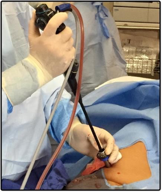
Fig. 6. Thoracoscope advanced into the pleural cavity under direct vision through the trocar. Reproduced from Alraiyes et al.¹⁶ under the terms of the Creative Commons Attribution-NonCommercial 3.0 License (CC BY-NC 3.0).
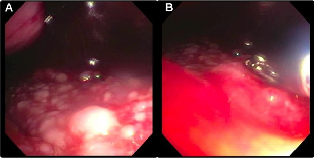
Fig. 7. (A) Multiple pleural densities visualised during thoracoscopy. (B) Thoracoscopic pleural biopsy obtained. Reproduced from Alraiyes et al.¹⁶ under the terms of the Creative Commons Attribution-NonCommercial 3.0 License (CC BY-NC 3.0).
5. Video-Assisted Thoracoscopic Surgery
Video-assisted thoracoscopic surgery (VATS) provides both diagnostic and therapeutic options in malignant pleural effusion (MPE), including pleural assessment, biopsy and talc pleurodesis. However, it is more invasive than medical thoracoscopy and generally requires general anaesthesia; it is therefore usually reserved for appropriately selected surgically fit patients. ²˒³
The AMPLE-3 trial compares IPC-based management with VATS pleurodesis in patients considered suitable for surgery, and its findings may help further define the role of surgical pleurodesis in MPE. ¹⁷
Choosing Between Definitive Pleural Interventions
In patients with recurrent symptomatic MPE who require definitive pleural intervention, the two principal treatment options are talc pleurodesis and an indwelling pleural catheter (IPC). Both can provide effective symptom relief and improvements in quality of life, but they differ in treatment burden, need for hospitalisation, suitability in non-expandable lung and the practical implications for patients. The choice should therefore be individualised according to lung expandability, prognosis, patient preference and local resources. Randomised trials including TIME2, AMPLE and, more recently, OPTIMUM have helped define the relative benefits and limitations of these approaches. ¹⁰˒¹¹˒¹⁸
The TIME2 trial (n=106) compared IPCs with chest tube insertion and talc slurry pleurodesis. There was no significant difference in dyspnoea at 42 days, the primary endpoint. However, patients managed with IPCs had significantly shorter initial hospitalisation (median 0 vs 4 days; p<0.001) and fewer subsequent pleural procedures (6% vs 22%; p=0.03). Adverse events were more frequent with IPCs (40% vs 13%; p=0.002), although most were non-serious catheter-related complications. ¹⁰
The AMPLE trial subsequently demonstrated that patients managed with IPCs spent fewer total days in hospital over 12 months than those undergoing talc pleurodesis (median 10 vs 12 days; p=0.03), while improvements in breathlessness and quality of life were comparable between groups. ¹¹
More recently, the OPTIMUM trial compared an outpatient IPC-based pathway, incorporating the option of talc instillation, with conventional inpatient chest drain insertion and talc pleurodesis. Health-related quality of life improved in both groups, with no significant difference in global health status at 30 days (mean difference 2.06; 95% CI −5.86 to 9.99; p=0.61), nor in breathlessness or chest pain. Outpatient IPC-based management was therefore not superior to standard inpatient care, supporting treatment selection based on patient preference, local expertise and healthcare resources. ¹⁸
Overall, the available evidence suggests that neither IPCs nor talc pleurodesis is universally superior. Talc pleurodesis may be preferred by patients with expandable lung who wish to avoid a long-term indwelling device and are suitable for a short inpatient admission. IPCs provide an effective ambulatory alternative and are particularly useful in patients with non-expandable lung, previous pleurodesis failure or a preference to minimise hospital stay. ²˒³
Shared decision-making is therefore central to definitive pleural management. Patients may prioritise different outcomes, including relief of breathlessness, avoidance of hospital admission, freedom from an indwelling device, convenience of treatment and maintenance of independence. ²˒³
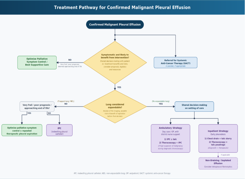
Fig 8. Suggested management pathway for symptomatic malignant pleural effusion. Author-generated flowchart informed by the British Thoracic Society Pleural Disease Guideline 2023 and the ATS/STS/STR Clinical Practice Guideline on malignant pleural effusion. ²˒³
Special Clinical Situations
(A) Non-expandable Lung
Non-expandable lung (NEL) refers to the inability of the lung to fully re-expand to the chest wall following pleural fluid drainage and affects up to approximately 30% of patients with MPE. ¹ It may result from visceral pleural restriction due to fibrous or malignant pleural involvement, or from endobronchial obstruction preventing full lung re-expansion.¹–³
Recognition of NEL is important because successful pleurodesis depends on close apposition of the visceral and parietal pleura following fluid evacuation. In patients with symptomatic MPE and NEL, indwelling pleural catheters (IPCs) are generally preferred because they can provide symptom control without requiring complete lung re-expansion.²˒³
Patients should, however, be counselled that breathlessness may not completely resolve despite adequate pleural drainage, reflecting the multifactorial nature of dyspnoea in advanced malignancy. Observational data suggest that NEL may not be associated with greater overall symptom burden or poorer health-related quality of life compared with expandable lung.¹⁹
(B) Septated Malignant Pleural Effusion
Septations may develop in malignant pleural effusion (MPE) through fibrin deposition and pleural adhesion formation, resulting in loculated collections that can impair drainage through a chest drain or indwelling pleural catheter (IPC). Residual pleural fluid may contribute to persistent breathlessness and reduce the likelihood of successful pleurodesis by preventing adequate apposition of the visceral and parietal pleura. Thoracic ultrasound is useful for identifying septations and assessing the extent and accessibility of residual pleural fluid. ¹˒²
Management of septated MPE can be challenging and should be individualised according to symptom burden, lung expandability, prognosis and previous pleural interventions. IPCs may be appropriate in selected patients, particularly in the presence of non-expandable lung or previous pleurodesis failure, although septations may themselves impair catheter drainage.²˒³
Evidence for intrapleural fibrinolytic therapy remains limited. In the TIME3 randomised controlled trial (n=71), intrapleural urokinase did not significantly improve dyspnoea (mean difference 3.8 mm; p=0.36) or pleurodesis failure (37% vs 32%; p=0.65) compared with placebo in patients with non-draining MPE. However, radiographic improvement in effusion size and a modest reduction in hospital stay of 1.6 days were observed; these secondary findings require further evaluation. ²⁰ Smaller studies and pooled evidence suggest that fibrinolytics may improve pleural drainage and, in selected patients, breathlessness or pleurodesis outcomes; however, the overall evidence base remains weak. ¹–³
Current BTS guidance therefore advises that intrapleural fibrinolytics may be considered in highly selected symptomatic patients with septated MPE. In patients with a septated MPE and an IPC, fibrinolytics may be used to improve drainage when flushing with normal saline or heparinised saline has been unsuccessful. ²
Surgical intervention may be considered for palliation in a small minority of patients with significantly septated MPE who remain symptomatic and have a favourable performance status and prognosis. Ultimately, management should prioritise symptom relief while balancing the potential benefit of further intervention against its procedural burden. ²˒³
Role of Systemic Anti-cancer Therapy
Systemic anti-cancer therapy (SACT) is an important component of malignant pleural effusion (MPE) management and should be considered according to tumour type, molecular characteristics, treatment responsiveness, performance status and prognosis. In treatment-sensitive malignancies, effective cancer-directed therapy may contribute to control of the underlying disease and pleural fluid accumulation; however, current evidence is insufficient to rely on SACT as a substitute for definitive pleural intervention in patients with symptomatic MPE.¹˒²
Definitive pleural intervention should therefore not routinely be deferred while awaiting a response to SACT. Pleural intervention and systemic therapy should instead be regarded as complementary: pleural procedures provide timely symptom relief and control of recurrent effusion, while SACT addresses the underlying malignancy. Close multidisciplinary collaboration between respiratory and oncology teams is essential to coordinate treatment according to individual patient priorities and goals of care. ¹˒²
Practical Considerations Across Healthcare Settings
Contemporary guidelines provide an evidence-based framework for the management of malignant pleural effusion (MPE). However, implementation of these recommendations may be influenced by the availability of pleural expertise, procedural resources and healthcare infrastructure. While indwelling pleural catheters (IPCs) have transformed ambulatory pleural care in many healthcare systems, access to catheter kits, community nursing support and drainage supplies may be limited in some settings.
Talc pleurodesis therefore remains an effective and widely applicable treatment option, particularly in centres where IPC services are unavailable or difficult to sustain. Similarly, repeated therapeutic aspiration may continue to have a role in carefully selected patients, especially when definitive interventions are not feasible or when treatment burden outweighs anticipated benefit. Clinical decision-making should balance contemporary evidence with pragmatic considerations, while maintaining a patient-centred approach and striving to provide the highest standard of care achievable within the local context.
Future Directions
Despite considerable advances in the management of malignant pleural effusion, several important clinical questions remain. Recent trials, including OPTIMUM¹⁸ and TACTIC¹⁵, continue to refine ambulatory pleural care and combined procedural strategies. The AMPLE-3 trial, comparing IPC-based management with video-assisted thoracoscopic surgery (VATS) pleurodesis in surgically fit patients, may further inform the optimal selection of definitive interventions. ¹⁷ Future research is likely to focus on improving patient selection, enhancing pleurodesis success, integrating patient-reported outcome measures and advancing personalised approaches to pleural care. ²¹
References
- Piggott, L.M., Hayes, C., Greene, J. and Fitzgerald, D.B. (2023) ‘Malignant pleural disease’, Breathe (Sheff.), 19(4), 230145. doi:10.1183/20734735.0145-2023.
- Roberts, M.E., Rahman, N.M., Maskell, N.A., Bibby, A.C., Blyth, K.G., Corcoran, J.P. et al. (2023) ‘British Thoracic Society Guideline for pleural disease’, Thorax, 78(11), pp. 1143–1156. doi:10.1136/thorax-2023-220304.
- Feller-Kopman, D.J., Reddy, C.B., DeCamp, M.M., Diekemper, R.L., Gould, M.K., Henry, T. et al. (2018) ‘Management of malignant pleural effusions. An official ATS/STS/STR clinical practice guideline’, American Journal of Respiratory and Critical Care Medicine, 198(7), pp. 839–849. doi:10.1164/rccm.201807-1415ST.
- Light, R.W., Macgregor, M.I., Luchsinger, P.C. and Ball, W.C. (1972) ‘Pleural effusions: the diagnostic separation of transudates and exudates’, Annals of Internal Medicine, 77(4), pp. 507–513. doi:10.7326/0003-4819-77-4-507.
- Arnold, D.T., De Fonseka, D., Perry, S., Morley, A., Harvey, J.E., Medford, A. et al. (2018) ‘Investigating unilateral pleural effusions: the role of cytology’, European Respiratory Journal, 52(5), 1801254. doi:10.1183/13993003.01254-2018.
- Porcel, J.M., Hernández, P., Martínez-Alonso, M., Bielsa, S. and Salud, A. (2015) ‘Accuracy of fluorodeoxyglucose-PET imaging for differentiating benign from malignant pleural effusions: a meta-analysis’, Chest, 147(2), pp. 502–512. doi:10.1378/chest.14-1243.
- Ost, D.E., Niu, J., Zhao, H., Grosu, H.B. and Giordano, S.H. (2018) ‘Quality gaps and comparative effectiveness of management strategies for recurrent malignant pleural effusions’, Chest, 153(2), pp. 438–452. doi:10.1016/j.chest.2017.08.026.
- Bhatnagar, R., Piotrowska, H.E.G., Laskawiec-Szkonter, M., Kahan, B.C., Luengo-Fernandez, R., Pepperell, J.C.T. et al. (2020) ‘Effect of thoracoscopic talc poudrage vs talc slurry via chest tube on pleurodesis failure rate among patients with malignant pleural effusions: the TAPPS randomized clinical trial’, JAMA, 323(1), pp. 60–69. doi:10.1001/jama.2019.19997.
- Fantin, A., Castaldo, N., Crisafulli, E., Sartori, G., Aujayeb, A., Vailati, P. et al. (2024) ‘The role of medical thoracoscopy with talc poudrage in spontaneous, iatrogenic, and traumatic pneumothorax: a prolonged experience of a tertiary care center’, Pulmonary Therapy, 10(3), pp. 347–362. doi:10.1007/s41030-024-00268-w.
- Davies, H.E., Mishra, E.K., Kahan, B.C., Wrightson, J.M., Stanton, A.E., Guhan, A. et al. (2012) ‘Effect of an indwelling pleural catheter versus chest tube and talc pleurodesis for relieving dyspnoea in patients with malignant pleural effusion (TIME2): a randomised controlled trial’, JAMA, 307(22), pp. 2383–2389. doi:10.1001/jama.2012.5535.
- Thomas, R., Fysh, E.T.H., Smith, N.A., Lee, P., Kwan, B.C.H., Yap, E. et al. (2017) ‘Effect of an indwelling pleural catheter versus talc pleurodesis on hospitalisation days in patients with malignant pleural effusion (AMPLE): a randomised clinical trial’, JAMA, 318(19), pp. 1903–1912. doi:10.1001/jama.2017.17426.
- Wahidi, M.M., Reddy, C., Yarmus, L., Feller-Kopman, D., Musani, A. and Shepherd, R.W. et al. (2017) ‘Randomized trial of pleural fluid drainage frequency in patients with indwelling pleural catheters: the ASAP Trial’, American Journal of Respiratory and Critical Care Medicine, 195(8), pp. 1050–1057. doi:10.1164/rccm.201607-1404OC.
- Muruganandan, S., Azzopardi, M., Fitzgerald, D.B., Shrestha, R., Kwan, B.C.H., Lam, D.C.L. et al. (2018) ‘Aggressive versus symptom-guided drainage of malignant pleural effusions via indwelling pleural catheters (AMPLE-2): an open-label randomised trial’, Lancet Respiratory Medicine, 6(9), pp. 671–680. doi:10.1016/S2213-2600(18)30288-1.
- Bhatnagar, R., Keenan, E.K., Morley, A.J. et al. (2018) ‘Outpatient talc administration by indwelling pleural catheter for malignant effusion’, New England Journal of Medicine, 378(14), pp. 1313–1322. doi:10.1056/NEJMoa1716883.
- Dipper, A., Sundaralingam, A., Hedley, E., Gudur, S., Mills, J., Sowden, S. et al. (2026) ‘Medical thoracoscopy with talc poudrage and indwelling pleural catheter insertion versus medical thoracoscopy with talc poudrage alone for patients with symptomatic malignant pleural effusion (TACTIC): a randomised, controlled phase 3 trial’, Lancet Respiratory Medicine, 14(4), pp. 341–349. doi:10.1016/S2213-2600(25)00409-6.
- Alraiyes, A.H., Dhillon, S.S., Harris, K., Kaphle, U. and Kheir, F. (2016) ‘Medical thoracoscopy: technique and application’, Pleura, 3, pp. 1–11. doi:10.1177/2373997516632752.
- Fitzgerald, D.B., Sidhu, C., Budgeon, C., Tan, A.L., Read, C.A., Kwan, B.C.H. et al. (2022) ‘Australasian Malignant Pleural Effusion (AMPLE)-3 trial: study protocol for a multi-centre randomised study comparing indwelling pleural catheter (±talc pleurodesis) versus video-assisted thoracoscopic surgery for management of malignant pleural effusion’, Trials, 23(1), 530. doi:10.1186/s13063-022-06405-7.
- Sivakumar, P., Fitzgerald, D.B., Ip, H., Rao, D., West, A., Noorzad, F. et al. (2024) ‘The impact of outpatient versus inpatient management on health-related quality of life outcomes for patients with malignant pleural effusion: the OPTIMUM randomised clinical trial’, European Respiratory Journal, 63(2), 2201215. doi:10.1183/13993003.01215-2022.
- Petersen, J.K., Fjaellegaard, K., Rasmussen, D.B., Alstrup, G., Høegholm, A., Sidhu, J.S. et al. (2024) ‘Patient-reported outcome measures in patients with and without non-expandable lung secondary to malignant pleural effusion—a single-centre observational study’, Diagnostics, 14(11), 1176. doi:10.3390/diagnostics14111176.
- Mishra, E.K., Clive, A.O., Wills, G.H., Davies, H.E., Stanton, A.E., Al-Aloul, M. et al. (2018) ‘Randomized controlled trial of urokinase versus placebo for nondraining malignant pleural effusion’, American Journal of Respiratory and Critical Care Medicine, 197(4), pp. 502–508. doi:10.1164/rccm.201704-0809OC.
- Gonnelli, F., Hassan, W., Bonifazi, M., Pinelli, V., Bedawi, E.O., Porcel, J.M. et al. (2024) ‘Malignant pleural effusion: current understanding and therapeutic approach’, Respiratory Research, 25(1), 47. doi:10.1186/s12931-024-02684-.
Author Information
Zalatt Pann Ei, Yadee Maung Maung Myint
1 Lister Hospital, East and North Hertfordshire Teaching NHS Trust, United Kingdom