Aetiology
Infectious, immune-mediated, and noninfectious triggers are all part of the genesis of GBS:
Infections
The most frequent antecedent.
- Campylobacter jejuni, hepatitis E virus, cytomegalovirus, Mycoplasma pneumoniae, Epstein-Barr virus, and HIV are the most frequent pathogens that cause seroconversion.
- Other links for infectious aetiology include varicella-zoster virus, Hemophilus influenzae, arboviruses (the Japanese encephalitis virus, dengue virus, chikungunya virus, and Zika virus).
- There is ongoing debate regarding the causal relationship between GBS and the COVID-19 virus.
Immunization
- The COVID-19 adenovirus vector vaccines may increase the risk of GBS, but not the messenger ribonucleic acid (mRNA) vaccines.
- A higher risk of GBS is linked to influenza vaccinations (especially 1976 swine influenza vaccine).
- The risk of GBS following vaccination with the recombinant zoster, hepatitis B, and human papillomavirus vaccines, the quadrivalent meningococcal diphtheria toxoid conjugate vaccine.
Despite the aforementioned hazards, most people now agree that vaccinations have advantages.
Noninfectious
The following are examples of potential noninfectious triggers:
- Surgical procedures
- Autoimmune diseases, such as systemic lupus erythematosus
- Trauma
- Drugs (such as type I interferons, tumor necrosis factor-α inhibitors, and immune
- checkpoint inhibitors)
- Malignant tumors, such as lymphomas
Epidemiology
The annual incidence of GBS is estimated to be between 1.1 and 1.8 per 100,000 person-years worldwide.
In children aged 0 to 15, the frequency is between 0.34 and 1.34 per 100,000 person-years.
Globally, the incidence varies, with rates ranging from 0.40 per 100,000 person-years in Brazil to 2.5 per 100,000 person-years in Bangladesh and Curaçao, and from 0.84 to 1.91 per 100,000 person-years in North America and Europe.
Infections precede about 70% of GBS cases. Men are impacted more frequently than women, with a ratio of around 1.5 : 1.
Pathophysiology and Pathogenesis
Immune Trigger and Molecular Mimicry
1.Preceding Infection:
- Most cases follow a respiratory or gastrointestinal infection (commonly Campylobacter jejuni, cytomegalovirus, Epstein-Barr virus, or influenza).
2.Molecular Mimicry:
- Microbial antigens share structural similarities with neural gangliosides or glycolipids found on peripheral nerves.
3.Antibody Production:
- The immune system generates cross-reactive immunoglobulin G (IgG) antibodies (such as anti-GM1) that target components of the host’s peripheral nerves.
Pathogenesis and Nerve Damage
1.Blood-Nerve Barrier Breakdown:
- Pro-inflammatory cytokines and immune activation facilitate the breakdown of the blood-nerve barrier.
2.Cellular Infiltration:
- T-lymphocytes, macrophages, and autoantibodies cross into the endoneurium, leading to inflammation and edema.
3.Complement Activation:
- Antibodies bind to neural targets and activate the classical complement pathway, leading to membrane attack complex (MAC) formation.
4.Macrophage Attack:
- Activated macrophages attach to and strip away myelin or directly damage axonal components.
Pathophysiologic Subtypes
1.Acute Inflammatory Demyelinating Polyradiculoneuropathy (AIDP):
- The immune response primarily targets and damages the myelin sheath and Schwann cells, causing slowed or blocked nerve conduction.
2.Acute Motor Axonal Neuropathy (AMAN) / Acute Motor and Sensory Axonal Neuropathy (AMSAN):
- The immune response directly attacks the axonal membrane, resulting in primary axonal degeneration and more severe structural damage.
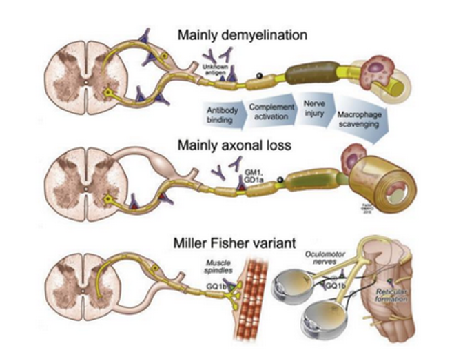
Fig. 1: Current understanding of Guillain-Barré syndrome pathogenesis and clinical variants. In demyelinating Guillain-Barré syndrome, unequivocal antigens have yet to be identified but are inferred by complement activation, myelin destruction, and cleanup by macrophages. In axonal and Miller Fisher variants, specific gangliosides (GM1, GD1a, GQ1b) are targeted by immunoglobulins and share antigenic epitopes with various bacterial and viral antigens. These antigenic targets are at nodal structures, at roots, and located at the end organs. In Miller Fisher syndrome, the GQ1b antigen also exists within the brain stem. In this variant, the macrophages clean up the axon debris and come in from the nodes.
Clinical Features
Classic sensorimotor GBS typically manifests about 10 days after the antecedent event.
The initial symptoms include lower back pain from nerve root inflammation and distal paresthesias.
Initial and Motor Features
1, Progressive Weakness
- Symmetrical weakness usually begins in the feet and legs before ascending to the arms, trunk, and face over hours, days, or weeks.
2.Flaccid Paralysis
- Deep tendon reflexes are reduced or absent in 90% of patients, and severe cases can lead to near-total flaccid paralysis.
3.Cranial Nerve Involvement
- Manifests as facial weakness, difficulty speaking, chewing, or swallowing (dysphagia), and eye movement problems (diplopia or blurred vision).
Sensory and Pain Features
1.Paresthesia
- Tingling, numbness, or a “pins and needles” sensation in the hands and feet frequently precedes or accompanies the onset of motor weakness.
2.Pain
- Deep, sharp, or shooting nerve pain commonly affects the back and legs in the early stages.
Autonomic and Critical Features
1.Respiratory Compromise
- Weakness of the chest muscles affects 1 in 3 people, leading to respiratory depression that may require mechanical ventilation.
2.Autonomic Dysfunction
- Fluctuations in blood pressure (hyper- or hypotension), irregular heart rhythms (arrhythmia or sinus tachycardia), and sluggish bowel or bladder function can occur.
Clinical state fluctuation
- Less than 5% of individuals experience recurrence, and the majority of GBS symptoms have a monophasic course.
- Treatment-related fluctuations, which are defined as up to two relapses within eight weeks of starting treatment, affect about 10% of patients.
- Reintroducing previously administered immunomodulatory medication usually resolves such treatment-related variations.
- The diagnosis of acute-onset chronic inflammatory demyelinating polyradiculoneuropathy (CIDP) should be taken into consideration in a subgroup of individuals who have three or more relapses or whose symptoms worsen after eight weeks.
- Subacute inflammatory demyelinating polyradiculoneuropathy is an intermediate subtype between GBS and CIDP that peaks between 4 and 8 weeks. Due to the possibility of future relapses and development to CIDP, these cases should be constantly followed.
GBS Subtypes
1.Electrophysiological Subtypes
- Acute Inflammatory Demyelinating Polyradiculoneuropathy (AIDP)
- Acute Motor Axonal Neuropathy (AMAN)
- Acute Motor-Sensory Axonal Neuropathy (AMSAN):
2.Clinical Variants
- Miller Fisher Syndrome (MFS)
- Pharyngeal-Cervical-Brachial (PCB) Variant
- Bickerstaff Brainstem Encephalitis (BBE)
- Acute Pan-dysautonomia
- Pure Sensory Variant
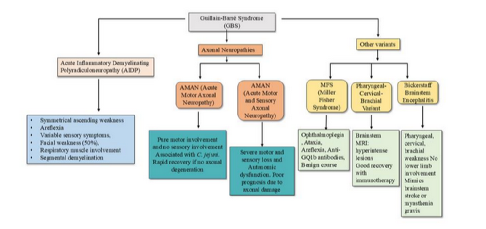
Fig. 2. Classification and Variants of Guillain-Barré Syndrome (GBS)
Evaluation
The diagnosis of GBS is primarily based on clinical history and neurologic examination.
Ancillary investigations such as cerebrospinal fluid (CSF) analysis and electrodiagnostic studies can support the diagnosis and help exclude mimics.
The Brighton criteria outline 4 levels of diagnostic certainty.
Key diagnostic features include clinical presentation, CSF findings, nerve conduction studies, and disease course. The diagnostic criteria have limitations and may miss variants of GBS.
Diagnostic criteria for Guillain–Barré syndrome
1.Features required for diagnosis
- Progressive bilateral weakness of arms and legs (initially only legs may be involved)
- Absent or decreased tendon reflexes in affected limbs (at some point in clinical course)
2.Features that strongly support diagnosis
- Progressive phase lasts from days to 4 weeks (usually <2 weeks)
- Relative symmetry of symptoms and signs
- Relatively mild sensory symptoms and signs (absent in pure motor variant)
- Cranial nerve involvement, especially bilateral facial palsy
- Autonomic dysfunction
- Muscular or radicular back or limb pain
- Increased protein level in cerebrospinal fluid (CSF); normal protein levels do not
rule out the diagnosis - Electrodiagnostic features of motor or sensorimotor neuropathy (normal electrophysiology in the early stages does not rule out the diagnosis)
Red flags for GBS diagnosis
- Increased numbers of mononuclear or polymorphonuclear cells in CSF (>50 × 106/l)
- Marked, persistent asymmetry of weakness
- Bladder or bowel dysfunction at onset or persistent during disease course
- Severe respiratory dysfunction with limited limb weakness at onset
- Sensory signs with limited weakness at onset
- Fever at onset
- Nadir <24 hours
- Sharp sensory level indicating spinal cord injury
- Hyper- reflexia or clonus
- Extensor plantar responses
- Abdominal pain
- Slow progression with limited weakness without respiratory involvement
- Continued progression for >4 weeks after start of symptoms
- Alteration of consciousness (except in Bickerstaff brainstem encephalitis)
Laboratory Investigations
The following laboratory tests help rule out other potential causes:
- Complete blood cell count
- Comprehensive metabolic profile with magnesium and phosphate levels
- Thiamine level
- Glycosylated hemoglobin
- Thyroid function testing
- Toxicology testing, if indicated
- Paraneoplastic panel (Especially in Hodgkin and non-Hodgkin lymphoma)
Anti-ganglioside antibody testing
It helps identify specific immune-mediated subtypes of Guillain-Barré syndrome (GBS).
It is not strictly required for a primary diagnosis.
1.Subtype Identification
- Particular antibodies correlate with distinct GBS variants such as anti-GQ1b and anti-GT1a IgG antibodies are strongly associated with Miller Fisher syndrome (MFS), while anti-GM1 and anti-GD1a are frequently seen in acute motor axonal neuropathy (AMAN).
2.Diagnostic Limitations
- Sensitivity ranges roughly from 35% to 60%, meaning a negative test result does not rule out GBS. Diagnosis remains primarily dependent on clinical presentation, nerve conduction studies, and cerebrospinal fluid (CSF) analysis.
Cerebrospinal fluid (CSF) analysis
It characteristically shows albumincytologic dissociation, which is an elevated total protein level with a normal or near-normal white blood cell count.
1.High Total Protein
- CSF protein levels are typically elevated (frequently >0.45 g/L to >0.55 g/L), reflecting inflammation and blood-nerve barrier dysfunction at the proximal nerve roots.
2.Normal Cell Count (Pleocytosis Absent)
- White blood cell counts usually remain normal (≤ 5 cells/μL).
- A marked increase in white blood cells suggests alternative diagnoses like infection or inflammation.
3.Timing Sensitivity
- CSF protein can be completely normal if the lumbar puncture is performed during the first week of symptom onset.
- Protein elevation is detected in over 50% to 70% of patients early on and rises to over 90% by the end of the second week.
4.Albumin Quotient (Q
- Measuring the CSF-to-serum albumin ratio helps assess blood-nerve barrier impairment independently of absolute protein reference shifts.
Clinical Utility CSF findings support a clinical diagnosis of GBS alongside nerve conduction studies.
The primary value of lumbar puncture is excluding mimics (such as infectious myelitis, poliomyelitis, or carcinomatous meningitis).
A normal protein level early in the disease course does not rule out GBS.
Electrophysiologic Studies
Nerve conduction studies (NCS) and needle electromyography (EMG) play a critical role in confirming the diagnosis, subtyping, and determining the prognosis of Guillain-Barré syndrome (GBS).
1.Early Indicators
- The earliest and most sensitive abnormality in GBS is the bilateral absence of the H-reflex and prolonged or absent F-wave latencies, which reflect proximal nerve root involvement before distal abnormalities appear.
2.General Nerve Conduction Changes
- Abnormalities in motor and sensory conduction appear in about 90% of patients, typically becoming fully apparent after the first week of symptoms.
- Findings include prolonged distal motor latencies, slowed conduction velocities, reduced compound muscle action potential (CMAP) amplitudes, and decreased sensory nerve action potential (SNAP) amplitudes.
3.Subtyping GBS
- Electrophysiological data help distinguish between the main subtypes of GBS.
4.Prognostic Value
Axonal Degeneration:
- Needle EMG findings of abundant spontaneous fibrillations and other signs of active axonal degeneration strongly associate with a poor prognosis and a slower rate of recovery.
Conduction Slowing
- Pure demyelinating features and the degree of initial conduction slowing generally do not correlate with long-term functional outcomes.
Neuroimaging
Neuroimaging is typically not required for diagnosing GBS.
Magnetic resonance imaging of the neuraxis with contrast enhancement may help support the diagnosis of GBS, especially in the presence of red flags.
Differential Diagnosis of GBS
Spinal Cord and Central Nervous System Disorders
- Acute myelopathy or transverse myelitis
- Acute cervical spinal cord injury or compression
- Bilateral strokes or brainstem lesions
Neuromuscular Junction and Toxic Disorders
- Myasthenia gravis
- Botulism
- Tick paralysis
- Neurotoxic fish or shellfish poisoning
Other Peripheral and Systemic Mimics
- West Nile virus infection
- Poliomyelitis
- Vasculitic or toxic neuropathies
- Porphyria polyneuropathy
Treatment / Management
Supportive care is the mainstay of GBS management.
ICU admission indications
The presence of one of the following must prompt consideration of admission to the intensive care unit:
- Dysautonomia (heart rate and blood pressure should be continually monitored)
- Bulbar dysfunction
- Severe or rapidly worsening weakness, especially of the neck and hip flexors
- Respiratory distress
Indications for Mechanical Intubation
Rapid disease progression, bulbar dysfunction, and weakening in the neck or hip flexors are the conditions that increase the likelihood of mechanical ventilation. Mechanical ventilation is indicated when signs of impending respiratory failure are present:
- Tachypnoea
- Accessory muscle use
- Inability to count to 15 or more during the expiratory phase of a single full capacity breath
- Diminished cough strength
- 20/30/40 rule: Vital lung capacity <20 mL/kg, or a maximal inspiratory pressure <30 cm H2O, or a maximal expiratory pressure <40 cm H2O
- Hypercarbia (partial pressure of arterial carbon dioxide >48 mm Hg) or hypoxemia (partial pressure of arterial oxygen <56 mm Hg)
Patients could have a sensation of breathlessness even in the presence of normal blood gas levels.
During intubation, depolarizing neuromuscular blockers like succinylcholine should be avoided since they can cause hyperkalemia. It is not advised to use noninvasive ventilation.
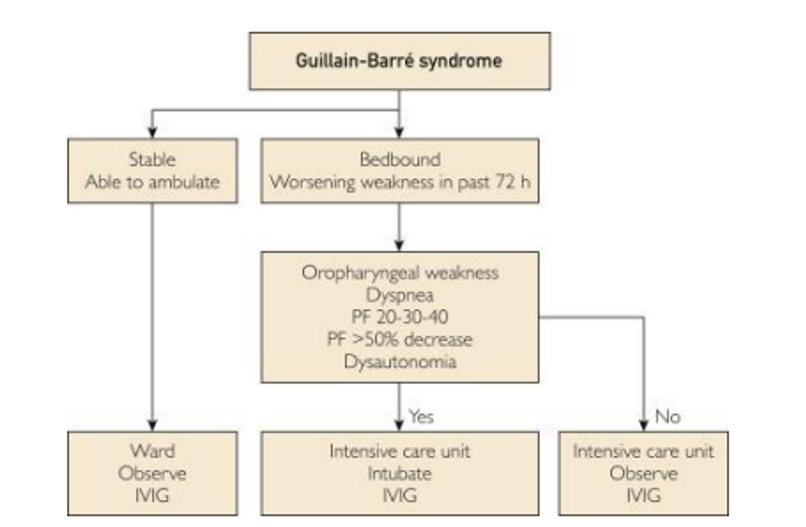
Fig. 3: Suggested triage criteria in hospitalized patients with Guillain-Barré syndrome.
IVIG = intravenous immunoglobulin; PF = pulmonary function; 20-30-40 = vital capacity decrease to 20 mL/kg, maximal inspiratory pressure decreases to −30 cm H2O, and maximal expiratory pressure decrease to 40 cm H2O.
When to start treatment
One or more of the following:
- Inability to walk >10 m independently
- Rapid progression of weakness
- Severe autonomic or swallowing dysfunction
- Respiratory insufficiency
Immunotherapy
In GBS, plasma exchange (PE) and intravenous immunoglobulin (IVIG) are equally beneficial.
Patient preference, geographical availability, cost, contraindications, and risk considerations should all be taken into consideration while deciding.
Because intravenous immunoglobulin (IVIG) is faster to administer and more tolerated, it is typically preferred.
These therapies can speed up healing, especially if started early, but they do not stop the disease from progressing or lessen the degree of nerve damage.
1.IVIG
- Over the course of two to five days, intravenous immunoglobulin is given at a dose of 2 g/kg. ((0.4 g/kg daily for 5 days)
- Transfusion reactions, headaches, aseptic meningitis, liver dysfunction, acute kidney damage from intravenous immunoglobulin products containing sucrose, thrombosis from hyperviscosity, and, in rare cases, anaphylaxis in individuals with IgA deficiency.
2.PE
- Each session of plasma exchange is given at a dose of 50 mL/kg plasma. (200–250/kg for 5 sessions)
- While serious instances might benefit from four sessions, milder cases might only need two.
- At least four sessions are necessary for severe cases; six sessions did not yield any extra benefits.
- Hypotension, transfusion responses, sepsis, thrombocytopenia, compromised coagulation parameters, hypocalcemia, and problems with intravenous access are among the complications.
- Patients with autonomic instability should generally avoid plasma exchange since significant fluid shifts can result in hypotension.
3. Factors considering if GBS has not improved with initial treatment
Approximately 40% of patients with Guillain–Barré syndrome (GBS) experiences no clinical improvement from standard immunotherapy (such as intravenous immunoglobulin or plasma exchange) after reaching a plateau, which typically occurs around 4 weeks.
- Plateau Phase
Many patients stop getting worse but fail to show immediate recovery after standard treatments. - Treatment-Related Fluctuations
Some patients improve initially and then get worse again, which may require repeating the immunotherapy. - Alternative Diagnoses
A lack of response can sometimes point to acute-onset chronic inflammatory demyelinating polyneuropathy (CIDP) rather than typical Guillain-Barré syndrome (GBS). - Ineffectiveness of Steroids
Clinical trials show that routine corticosteroids do not help standard GBS and can lead to worse outcomes. - Rehabilitation
Physical therapy, occupational therapy, nutritional support, and speech-language pathology must be involved early.
Indications for Tracheostomy
- Early tracheostomy is expected in elderly patients with preexisting pulmonary disease and in those with severe axonal involvement by electromyographic criteria.
- If patients are intubated for oropharyngeal weakness and the disease continues to progress, tracheostomy should be performed also to avoid ventilator-associated pneumonia.
- Postponing tracheostomy is an option in patients with dubious reasons for intubation or improving pulmonary function test result.
- Those postponements can be accomplished in only 1 in 5 patients at the most.
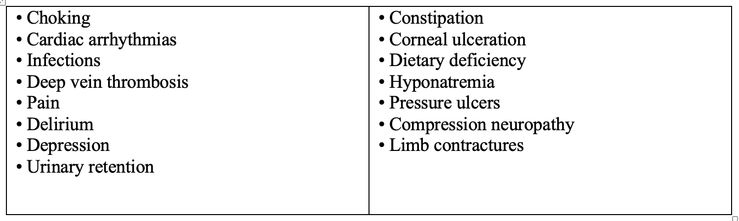
Complications
Prognosis
Long-term results in GBS are generally positive.
Within a year, over 50% of patients make a full recovery. At six months and twelve months following immunotherapy, 77% and 81% of patients were able to walk on their own.
- Although some patients do not fully recover, axonal GBS may continue to improve after a year.
- Recovery for GBS variants differ from typical GBS type.
- Weakness, neuropathic pain, exhaustion, and psychological symptoms like depression and grief are common signs of long-term disability.
- The overall mortality rate is between 3% and 7%, and it is roughly 20% for patients who need mechanical ventilation.
- Prognosis does not appear to be impacted by sex.
- Higher mortality is indicated by advanced age, related comorbidities, severe illness, heart and lung problems, mechanical ventilation, and systemic infection.
- Axonal electrophysiological subtypes and antecedent C jejuni infection, particularly if accompanied by diarrhea, are additional indicators of a poor outcome.
- The most frequent causes of death are infections, acute respiratory distress syndrome, and pulmonary embolism. These incidents can happen both during the acute and recovery phases, highlighting the necessity of ongoing supportive care.
Summary
- Classic Guillain–Barré syndrome (GBS) is an acute- onset ascending sensorimotor neuropathy, but the disease can present atypically or as a clinical variant.
- Abnormal results in electrophysiological studies and a combination of an increased protein level and normal cell count in cerebrospinal fluid are classic features of GBS, but patients with GBS can have normal results in both tests, especially early in the disease course.
- Respiratory function should be monitored in all patients as respiratory failure can occur without symptoms of dyspnea.
- Intravenous immunoglobulin and plasma exchange are equally effective in treating GBS; no other treatments have been proven to be effective.
- The efficacy of repeat treatment in patients who have shown insufficient clinical response is uncertain; but this practice is common in patients who show deterioration after an initial treatment response.
- Clinical improvement is usually most extensive in the first year after disease onset and can continue for >5 years.
- Clinicians, nurses, occupational therapists, physical therapists, and speech therapists should all be involved in the interprofessional management of complications.
Further Reading
- Hughes RA, Pritchard J, Hadden RD. Pharmacological treatment other than corticosteroids, intravenous immunoglobulin and plasma exchange for Guillain-Barré syndrome. Cochrane Database Syst Rev. 2013 Feb 28. 2:CD008630.
- Kushnir M, Klein C, Pollak L, Rabey JM. Evolving pattern of Guillain-Barre syndrome in a community hospital in Israel. Acta Neurol Scand. 2008 May. 117(5):347-50.
- Li H, Yuan J. Miller Fisher syndrome: toward a more comprehensive understanding. Chin Med J (Engl). 2001 Mar. 114(3):235-9.
- McGillicuddy DC, Walker O, Shapiro NI, Edlow JA. Guillain-Barré syndrome in the emergency department. Ann Emerg Med. 2006 Apr. 47(4):390-3.
- Raphaël JC, Chevret S, Hughes RA, Annane D. Plasma exchange for Guillain-Barré syndrome. Cochrane Database Syst Rev. 2012 Jul 11. 7:CD001798.
- Rudolph T, Larsen JP, Farbu E. The long-term functional status in patients with Guillain-Barré syndrome. Eur J Neurol. 2008 Dec. 15(12):1332-7.
- Seneviratne U. Guillain-Barré syndrome. Postgrad Med J. 2000 Dec. 76(902):774-82.
- Toscano G, Palmerini F, Ravaglia S, et al. Guillain-Barré Syndrome Associated with SARS-CoV-2. N Engl J Med. 2020 Apr 17.
- van der Meché FG, Schmitz PI. A randomized trial comparing intravenous immune globulin and plasma exchange in Guillain-Barré syndrome. Dutch Guillain-Barré Study Group. N Engl J Med. 1992 Apr 23. 326(17):1123-9.
- van Doorn PA. What’s new in Guillain-Barré syndrome in 2007-2008?. J Peripher Nerv Syst. 2009 Jun. 14(2):72-4.
- Winer JB. Treatment of Guillain-Barré syndrome. QJM. 2002 Nov. 95(11):717-21.
Author Information
Dr. Thar Thar Oo
Senior Consultant Neurologist