Introduction
Coal workers’ pneumoconiosis (CWP) is an occupational lung disease resulting from prolonged inhalation of respirable coal mine dust. It forms part of the broader spectrum of coal mine dust lung disease, which includes simple CWP, progressive massive fibrosis, chronic bronchitis, emphysema, airflow obstruction, and dust-related diffuse fibrosis 1,2. The risk of disease is related primarily to cumulative dust exposure, although the intensity of exposure and the silica content of inhaled dust are also important determinants 1–3.
The diagnosis of CWP is generally based on a compatible occupational exposure history together with characteristic radiological findings. Lung biopsy is not routinely required but may be useful when the diagnosis remains uncertain or when competing infectious, malignant, or other fibrotic lung diseases cannot be confidently excluded 1,2.
Diagnosing acute/chronic pulmonary disease in patients with advanced human immunodeficiency virus (HIV) infection can be particularly challenging. Pulmonary tuberculosis, Pneumocystis jirovecii pneumonia (PJP), bacterial pneumonia, and other opportunistic infections frequently dominate the initial differential diagnosis. Consequently, coexisting non-infectious disorders, including occupational lung disease, may be overlooked.
We report a diagnostically challenging case of biopsy-confirmed CWP in a patient with advanced HIV infection whose persistent hypoxaemic respiratory illness was initially attributed to pulmonary infectious and opportunistic causes.
Case Report
A 48-year-old married man from Danuphyu, Myanmar, was admitted to Tropical and Infectious Diseases Department of Yangon General Hospital on 25 February 2026 with progressive exertional dyspnoea for two months. The dyspnoea was associated with productive cough with whitish sputum. He denied fever, night sweats, significant weight loss, haemoptysis, wheezing, chest pain, orthopnoea, or peripheral oedema.
He had been diagnosed with HIV infection in 2014 and commenced antiretroviral therapy (ART). He defaulted ART in 2021 but restarted treatment in 2022. In April 2024, he had been evaluated for chronic respiratory symptoms and abnormal chest radiographic findings and was treated for presumed pulmonary tuberculosis, completing a full course of anti-tuberculosis therapy. Following completion of treatment, he was lost to follow-up because of financial difficulties.
He has a lifelong non-smoker and did not consume alcohol.
On examination, he was conscious and oriented but appeared mildly malnourished and dyspnoeic. There was no pallor, cyanosis, clubbing, peripheral oedema, or significant lymphadenopathy. Respiratory examination revealed mildly reduced bilateral chest expansion and bilateral fine end-inspiratory crepitations, predominantly over the upper lung zones. No wheeze or pleural rub was detected.
His blood pressure was 100/70 mmHg and pulse rate was 80 beats/min. Oxygen saturation was 86% on room air, improving to 95% with oxygen at 3 L/min. Cardiovascular, neurological, and abdominal examinations were unremarkable.
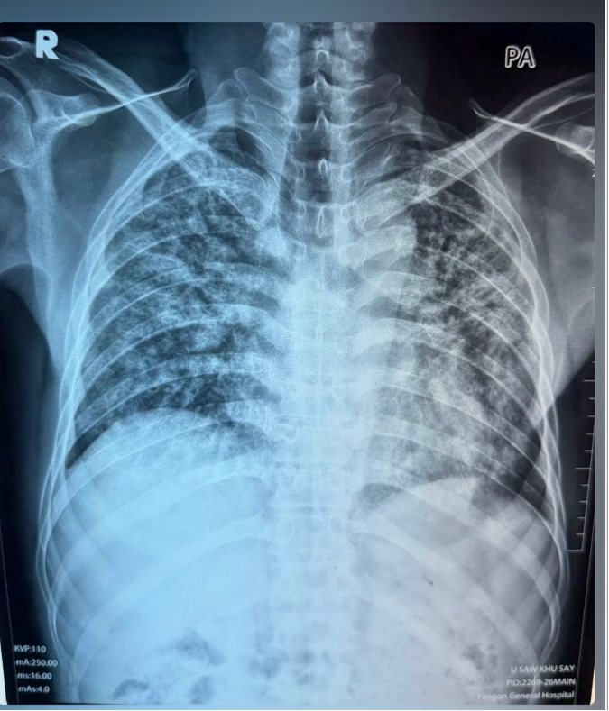
Fig. 1. Posteroanterior chest radiograph showing bilateral diffuse reticulonodular/interstitial pulmonary opacities, more prominent in the upper and mid lung zones.
The CD4 count was 87 cells/µL, while HIV viral load was suppressed at <40 copies/mL. Sputum Xpert MTB/RIF was negative, and urinary lipoarabinomannan was also negative. Sputum bacterial culture isolated Escherichia coli susceptible to several antimicrobial agents.
In view of the bacterial culture result, intravenous meropenem was administered. However, there was no significant clinical improvement. Because of the low CD4 count and persistent hypoxaemic respiratory illness, PJP was also clinically considered. He received therapeutic trimethoprim-sulfamethoxazole together with corticosteroids for three weeks, but his respiratory symptoms persisted.
Sputum fungal examination demonstrated budding yeast with pseudohyphae, and culture isolated Candida albicans. In the setting of advanced immunosuppression, pulmonary fungal infection was considered and liposomal amphotericin B was administered.
During hospitalization, the patient became increasingly immobile with persistent hypoxaemia and tachycardia. His D-dimer was approximately five times the upper limit of normal, with a Wells score of 3. Computed tomography pulmonary angiography (CTPA) was therefore performed on 16 March 2026. There was no evidence of pulmonary thromboembolism. However, CTPA demonstrated extensive pulmonary abnormalities including pulmonary fibrosis and traction bronchiectasis involving both upper lobes, mild cardiomegaly, pulmonary arterial hypertension, and mediastinal lymphadenopathy.
Persistent respiratory impairment despite treatment directed against the suspected infectious conditions prompted reconsideration of the diagnosis. A more detailed occupational history was subsequently obtained, revealing that the patient had worked as a coal miner at Mine Li Coal Mine for seven years.
High-resolution computed tomography (HRCT) of the chest was performed on 18 March 2026. It demonstrated fibrotic strands in both lungs, predominantly affecting the upper lobes, with volume loss of both upper lobes and the right middle lobe. Traction bronchiectasis was present bilaterally, predominantly in the upper lung fields. Small cystic lesions were also noted in both lungs.
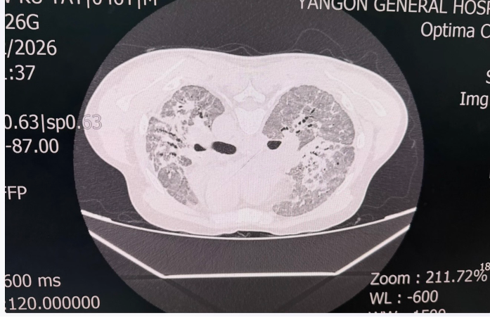
Fig 2. Axial HRCT chest image demonstrating bilateral upper-lobe-predominant fibrotic change with architectural distortion and traction bronchiectatic changes.
Although the occupational exposure raised the possibility of CWP, the imaging findings were not considered sufficiently specific to distinguish pneumoconiosis from previous tuberculosis, chronic pulmonary infection, or other causes of fibrotic lung disease. The diagnostic uncertainty was particularly important in view of the patient’s advanced HIV infection and previous treatment for pulmonary tuberculosis.
A CT-guided lung biopsy was therefore performed on 22 March 2026. Histopathological examination demonstrated black particles engulfed by alveolar macrophages with adjacent collagenous nodules. There was no evidence of tuberculosis or malignant change. The pathological findings were reported as fibrotic lung with anthracosis compatible with pneumoconiosis.
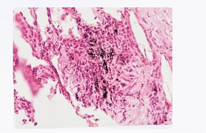
Fig. 3. Lung biopsy histopathology showing black carbonaceous pigment within alveolar macrophages with adjacent fibrotic/collagenous tissue, consistent with anthracosis and coal workers’ pneumoconiosis.
The combination of occupational coal mine dust exposure, upper-lobe-predominant fibrotic lung disease, and characteristic histopathological findings established the diagnosis of coal workers’ pneumoconiosis.
The patient was managed with supplemental oxygen, bronchodilator therapy, chest physiotherapy, and continuation of ART. Further exposure to coal mine dust and other respiratory hazards was discouraged.
Discussion
Coal mine dust lung disease encompasses several pulmonary disorders resulting from occupational exposure to respirable coal mine dust. Classical CWP is characterized by the deposition of coal dust within the lungs and the development of coal macules and nodules, while more advanced disease may progress to extensive fibrosis or progressive massive fibrosis 1–3.
The pathological hallmark of CWP is the accumulation of carbonaceous dust-laden macrophages, particularly around respiratory bronchioles, accompanied by varying degrees of reticulin and collagen fibrosis 2,3. In the present case, black pigment-laden alveolar macrophages with adjacent collagenous nodules provided important histopathological evidence supporting the diagnosis.
The duration of occupational exposure in our patient was seven years. Although CWP is commonly associated with longer cumulative exposure, shorter-duration exposure does not exclude the diagnosis when dust concentrations are high 1,2. Exposure intensity, working conditions, ventilation, proximity to dust-generating activities, and the crystalline silica content of coal mine dust may substantially influence the risk of disease 1–3.
The major diagnostic challenge in this patient was the presence of advanced HIV infection with a CD4 count of 87 cells/µL. In such patients, pulmonary tuberculosis, PJP, bacterial pneumonia, and other opportunistic infections appropriately constitute important initial diagnostic considerations. Previous pulmonary tuberculosis further complicated interpretation of the upper-lobe fibrosis and traction bronchiectasis.
The microbiological findings also contributed to the initial emphasis on infection. E. coli was isolated from sputum, and PJP was treated clinically because of the patient’s severe immunosuppression. Nevertheless, the absence of meaningful clinical improvement despite directed antimicrobial treatment suggested that infection alone could not explain the underlying chronic pulmonary process.
The isolation of Candida albicans from sputum should be interpreted cautiously. Candida species commonly colonize the respiratory tract, and their isolation from respiratory secretions generally does not establish invasive Candida pneumonia 4. Histopathological evidence of tissue invasion is usually necessary for definitive diagnosis 4. But anti fungal treatment was started in this patient because of severely immunocompromised with CD4 count 87 cells / ml.
The turning point in this case was the recognition of the patient’s occupational history. Once his seven-year history of coal mining was identified, an occupational cause for the upper-lobe fibrotic abnormalities became an important consideration. This highlights the importance of obtaining a detailed occupational history in every patient presenting with unexplained chronic respiratory disease.
HRCT is more sensitive than plain chest radiography for characterizing emphysema, fibrosis, and other manifestations of coal mine dust lung disease 1. However, imaging findings may overlap with tuberculosis, other granulomatous diseases, malignancy, and other fibrotic lung disorders. Lung biopsy is therefore not routinely required for CWP but may be valuable when significant diagnostic uncertainty remains 1,2.
In the present case, the combination of advanced HIV infection, previous pulmonary tuberculosis, microbiological findings, and non-specific fibrotic changes on HRCT made a purely radiological diagnosis difficult. Histopathological examination demonstrating anthracotic pigment within alveolar macrophages and adjacent collagenous nodules was therefore pivotal in establishing the diagnosis.
There is currently no specific pharmacological therapy that reverses established CWP. Management is primarily supportive and includes prevention of further dust exposure, treatment of associated airflow obstruction and respiratory infections, appropriate vaccination, pulmonary rehabilitation, and supplemental oxygen when indicated 1,2. Importantly, CWP may continue to progress even after occupational exposure has ceased, emphasizing the importance of continued clinical and respiratory follow-up 1.
This case illustrates an important diagnostic principle in immunocompromised patients: although infectious and opportunistic diseases should be actively investigated, persistent symptoms despite appropriate therapy should prompt reconsideration of the diagnosis and evaluation for non-infectious causes. A carefully obtained occupational history may provide the decisive diagnostic clue.
Conclusion
Coal workers’pneumoconiosis should be considered in patients with chronic respiratory symptoms and fibrotic lung abnormalities when there is a relevant history of coal mine dust exposure, even when the duration of occupational exposure is relatively short.
In patients with advanced HIV infection, pulmonary tuberculosis and opportunistic infections may dominate the initial diagnostic approach and obscure coexisting non-infectious pulmonary disease. Failure to respond to appropriately directed antimicrobial therapy should therefore prompt reassessment of the original diagnosis.
This case emphasizes the importance of detailed occupational history-taking and demonstrates the value of histopathological confirmation when clinical, radiological, and microbiological findings remain inconclusive.
Conclusion
- Petsonk EL, Rose C, Cohen R. (2013). Coal mine dust lung disease: new lessons from an old exposure. Am J Respir Crit Care Med. 187(11):1178-1185.
- Cohen RA, Petsonk EL, Rose C, et al. (2016). Lung pathology in U.S. coal workers with rapidly progressive pneumoconiosis implicates silica and silicates. Am J Respir Crit Care Med. 193(6):673-680.
- Vallyathan V, Brower PS, Green FHY, Attfield MD. (1996). Radiographic and pathologic correlation of coal workers’ pneumoconiosis. Am J Respir Crit Care Med. 154(3):741-748.
- Pappas PG, Kauffman CA, Andes DR, et al. (2016). Clinical practice guideline for the management of candidiasis: 2016 update by the Infectious Diseases Society of America. Clin Infect Dis. 62(4).
Author Information
Aye-Mya-Theingi-Win1, Khin-Rupar-Ko2, Aye-Aye-Win3, May-Zabe4, Nyunt Thein5
- Senior Consultant physician, Tropical and Infectious Diseases Department, Yangon General Hospital
- Professor, Tropical and Infectious Diseases Department, Yangon General Hospital
- Associate Professor, Tropical and Infectious Diseases Department, University of Medicine (1), Yangon
- Senior Consultant Physician, Tropical and Infectious Diseases Department, Yangon General Hospital
- Senior Consultant Physician, Former Head of Department of Medicine, Emeritus Professor of Medicine, University of Medicine (1), Yangon